Carotid Ultrasound
Carotid (ka-ROT-id) ultrasound, also called Doppler ultrasound or Carotid duplex ultrasound, is a painless and harmless test that uses high-frequency sound waves to create pictures of the insides of the two large arteries in your neck. These arteries, called carotid arteries, supply your brain with oxygen-rich blood. You have one carotid artery on each side of your neck.
Carotid ultrasound shows whether a substance called plaque (plak) has narrowed your carotid arteries. Plaque is made up of fat, cholesterol, calcium, and other substances found in the blood. Plaque builds up on the insides of your arteries as you age. This condition is called Carotid Artery Disease.
A carotid ultrasound can show whether plaque buildup has narrowed one or both of your carotid arteries and reduced blood flow to your brain. If plaque has narrowed your carotid arteries, you may be at risk of having a stroke. That risk depends on how much of your artery is blocked and how much blood flow is restricted. To reduce your risk for stroke, your doctor may recommend medical or surgical treatments to reduce or remove the plaque buildup in your carotid arteries.
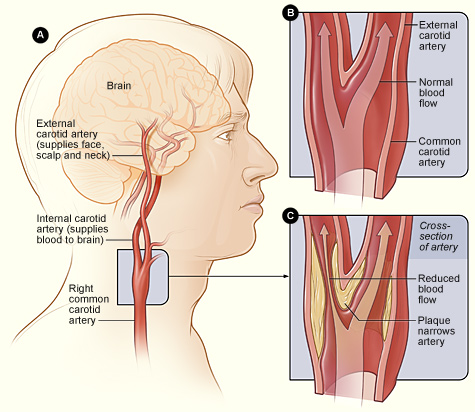
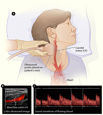
Figure A shows how the ultrasound probe (transducer) is placed over the carotid artery. Figure B is a color ultrasound image showing blood flow (the red color in the image) in the carotid artery. Figure C is a waveform image showing the sound of flowing blood in the carotid artery.

Candidates for Carotid Ultrasound
Carotid ultrasound checks for plaque buildup in the carotid arteries. Plaque can narrow or block your carotid arteries, preventing oxygen-rich blood from reaching your brain. Your doctor may recommend a carotid ultrasound if you:
- Have had a stroke or mini-stroke recently. During a mini-stroke, you may have some or all of the symptoms of a stroke. However, the symptoms usually go away on their own within 24 hours.
- Have an abnormal sound in your carotid artery called a carotid bruit (broo-E). Your doctor can hear a carotid bruit with the help of a stethoscope put on your neck over the carotid artery. A bruit may suggest a partial blockage in your carotid artery that could lead to a stroke.
Your doctor may also recommend a carotid ultrasound if he or she suspects you may have:
- Blood clots that can slow blood flow in your carotid artery.
- A split between the layers of your carotid artery wall that weakens the wall or reduces blood flow to your brain.
A carotid ultrasound may also be done to see whether carotid artery surgery, also called carotid endarterectomy (END-ar-ter-EK-to-me), has restored normal blood flow through your carotid artery.
If you had a procedure called carotid stenting, you may have a carotid ultrasound afterward to check the position of the stent put in your carotid artery. (The stent, a small mesh tube, helps prevent the artery from becoming narrowed or blocked again.)
Sometimes carotid ultrasound is used as a preventive screening test in people who have medical conditions that increase their risk of stroke, including high blood pressure and diabetes. People who have these conditions may benefit from having their carotid arteries checked regularly, even if they show no signs of plaque buildup.
Description of the Procedure
Carotid ultrasound usually is done in a doctor's office or hospital. The test is painless and often doesn't take more than 30 minutes.
The ultrasound machine includes a computer, a video screen, and a transducer. A transducer is a hand-held device that sends and receives ultrasound waves into and from the body.
You will lie on your back on an exam table for the test. Your technician or doctor will put a gel on your neck where your carotid arteries are located. This gel helps the ultrasound waves reach the arteries better.
Your technician or doctor will put the transducer against different spots on your neck and move it back and forth. The transducer gives off ultrasound waves and detects their echoes after they bounce off the artery walls and blood cells. Ultrasound waves can't be heard by the human ear.
A computer uses the echoes to create and record pictures of the insides of the arteries (usually in black and white) and your blood flowing through them (usually in color; this is the Doppler ultrasound). A video screen displays these live images for your doctor to review.
Carotid Ultrasound
Figure A shows how the ultrasound probe (transducer) is placed over the carotid artery. Figure B is a color ultrasound image showing blood flow (the red color in the image) in the carotid artery. Figure C is a waveform image showing the sound of flowing blood in the carotid artery.